
Digital Dental X Rays in Las Vegas: What to Know
You're settling into the dental chair for a new-patient exam in Las Vegas when the team asks to take X-rays. You might wonder whether they're necessary, how much radiation they involve, or whether your old images could be used instead. Those are reasonable questions, and you deserve a clear answer before imaging begins.
Digital dental X-rays help a dentist look for problems that may not be visible during an exam or cleaning, such as decay between teeth, changes around a tooth's root, or bone loss. At Aspiring Smiles Dental and Braces, imaging can support decisions about preventive care, restorative dentistry, cosmetic treatment, and urgent concerns. The right images depend on your symptoms, oral health, and treatment needs, not just on a routine schedule.
Why Your Dentist Recommends Digital Dental X Rays
A visual exam gives your dentist important information, but it can't show every surface between teeth or the structures below the gums. If you've come in with tooth pain, for example, an image may help the dentist assess whether the concern involves decay, a root, or surrounding bone. During a new-patient exam, images can also help establish a useful record when there aren't recent X-rays available.
That doesn't mean every visit automatically calls for imaging. A dentist should consider your health history, symptoms, previous images, and the reason for the appointment before recommending X-rays. If you're visiting an emergency dentist because of pain or a cracked tooth, the team may focus on the area involved. For a routine visit, the dentist may decide that existing images are still useful.
What images can help reveal
Dental X-rays can contribute to decisions about whether to monitor a finding, provide preventive care, or discuss treatment. A small area of decay between teeth may be difficult to spot by looking into the mouth alone. Images can also help evaluate bone levels and roots when a dentist is considering restorative work, tooth extraction, or dental implants.
For cosmetic dentistry, imaging is one piece of a broader evaluation. A dentist may need to understand the health of teeth and supporting structures before discussing options such as veneers or crowns. X-rays don't show every detail relevant to a smile plan, but they can help identify issues that need attention first.
Before imaging: Ask what the X-ray is intended to help the dentist assess and whether prior images could answer the same question.
If you're comparing a dentist near me in Las Vegas, NV, it's fair to ask how the office decides when images are needed. The aim is to gather information that supports care designed for you, not to take images just because equipment is available.
How Digital Dental X Rays Actually Work
A film X-ray is a little like taking a photograph that needs to be developed before you can see it. Digital imaging uses an electronic receptor or a reusable plate to capture the X-ray information, then displays the image on a computer. That lets the dentist review it with you without sending film away for chemical processing.
Sensors, plates, and wider views
A solid-state sensor is a small electronic device placed in your mouth to capture an image. Two common types are CCD and CMOS sensors. You can think of them as digital camera sensors: they turn the image information into a file the computer can display.
A photostimulable phosphor (PSP) plate is closer to a reusable film substitute. It records the image and then goes through a scanner before the image appears on screen. The plate and sensor systems work differently, but both create digital images for the dental team to review.
A panoramic unit captures a broad view of the teeth and jaws rather than a close-up of a small area. A cone beam computed tomography (CBCT) scan produces three-dimensional information for cases where the dentist needs a more detailed view. For a closer look at that imaging option, read about 3D cone beam dental scans in Las Vegas.
| Type | What It Shows | Typical Use |
|---|---|---|
| Intraoral sensor | A focused view of selected teeth | Checking a specific area, such as teeth or roots |
| PSP plate | A focused digital image after scanning | Taking targeted dental images |
| Panoramic X-ray | A broad view of both jaws and teeth | Reviewing a wider area |
| CBCT scan | Three-dimensional details of teeth and surrounding structures | Planning when a 3D view is clinically useful |
Once the image is displayed, the dentist may enlarge it or adjust brightness and contrast. These tools can make details easier to examine, but they don't turn an unclear image into a diagnosis. Your dentist still considers the image alongside your symptoms, exam, and dental history.
Digital X Rays Compared to Traditional Film
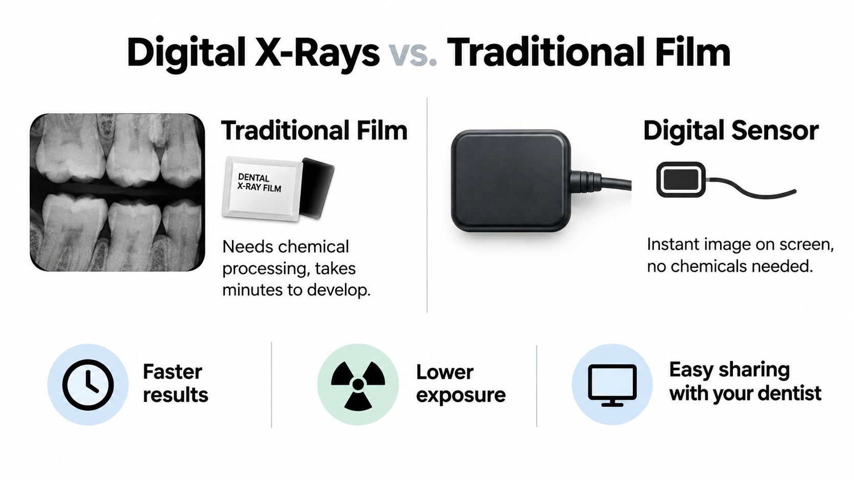
If you had dental X-rays years ago, you may remember biting down on a small film packet and then waiting while it was developed. Film needs chemical processing before anyone can look at it. Digital imaging skips that step entirely: the image appears on a screen within moments, so the dentist can pull it up beside your chair and walk you through what they see during the same visit.
The differences continue after the appointment. A digital file stays in your chart, can be reviewed side by side at future visits, and travels electronically if your care involves another provider, whether that is a specialist across town or a new Las Vegas office. Digital systems also skip the chemical processing that film requires.
A more comfortable process still depends on fit
Convenience does not change how the receptor feels in your mouth. A digital sensor can seem firm or bulky, especially with a small mouth or a sensitive gag reflex. If X-rays have been uncomfortable for you before, say so before the image is taken. The team can explain the positioning first, adjust how the receptor is placed, or pause so you can take a breath.
One honest caveat about the technology: a lower dose per image is only part of the picture. When imaging becomes quick and easy, a practice can end up taking more images, and extra radiographs can eat into the dose savings digital systems offer. Public-health guidance likewise emphasizes selecting patients and examinations thoughtfully instead of imaging on a fixed schedule. Before your next X-ray, you can ask how the office decides a new image is needed and whether your existing images get reviewed first. Both are fair questions, and a good team will answer them plainly.
Radiation Safety and Realistic Exposure Numbers
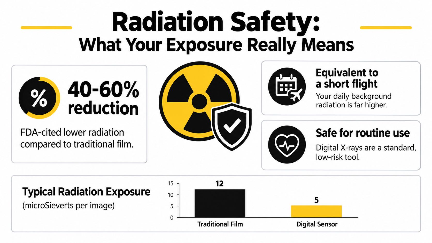
A patient in the chair often asks some version of the same question: how much radiation am I getting? The honest answer starts with the sensor itself. Digital receptors respond to X-rays more efficiently than film, so a team using appropriate equipment settings can capture an image with less exposure. Federal guidance on dental radiographic exams, cited earlier in this article, frames that switch as an opportunity to reduce patient dose by roughly 40% to 60%. A review of digital dental radiography reports an even wider gap in some comparisons, finding digital systems may require approximately 90% less radiation than E-speed film. Treat those figures as a range of possibilities, not a promise about your personal dose.
Even one image type has no single dose. In a study of five digital panoramic units, effective doses ran from 4.7 to 14.9 microsieverts per exposure. Same kind of image, about a threefold spread. Machine design and the protocol the operator selects account for much of that difference, which is why the word "digital" alone cannot tell you what you will receive.
Why the number of images matters
Radiation safety in dentistry follows a principle called ALARA: keep exposure as low as reasonably achievable while still capturing the diagnostic information your care requires. A lower dose per image helps only when the total count stays reasonable. Patient selection, careful positioning, correct settings, and disciplined retake decisions each carry part of that responsibility.
The American Dental Association says there's no universal schedule for dental X-rays. Timing depends on your age, current oral health, disease risk, and any signs or symptoms, according to the ADA's guidance on dental X-rays and radiographs. The ADA also recommends reusing prior images when they remain diagnostically useful, and it cautions that digital equipment still demands proper technique.
That gives you a practical script for a Las Vegas office. Before anyone takes an X-ray, ask whether your previous images were reviewed, why a new one is needed, how the team limits retakes, and whether computer-assisted reading plays a role in reviewing what the sensor captures. A good team answers plainly, and those answers reveal more about a practice's safety culture than any single number on a brochure.
What to Expect During Your Imaging Appointment
Imaging usually starts with a short conversation. At a new-patient exam, or during an emergency visit for pain, the dental team should ask about your concern and review what records you have. The dentist then decides which images, if any, could help answer a clinical question.
For an intraoral image, the hygienist or dentist places a sensor or plate inside your mouth and positions it near the area being examined. You'll be asked to hold it in place briefly while the image is captured. If the position feels uncomfortable or you have a strong gag reflex, say so. The team can explain what they're doing and make adjustments when possible.
From image to explanation
The team may use a protective apron or thyroid collar according to the situation and the practice's protocols. Proper beam alignment matters, too. Rectangular collimation narrows the X-ray beam to the area that needs to be examined, rather than covering a wider field without need.
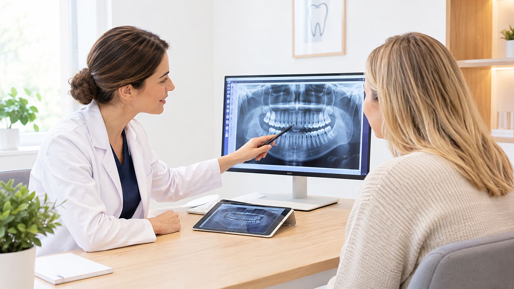
After the image appears on screen, the dentist can review it, adjust how it's displayed, and compare it with earlier images if available. You should have an opportunity to ask what the image shows and how it relates to your symptoms or treatment plan. Images help inform a clinical decision, but the dentist interprets them alongside your exam and health history.
You can bring a short list of questions to your appointment:
- Purpose: What concern is this image meant to evaluate?
- Prior records: Can you use an image from my previous dentist?
- Retakes: What would make a retake necessary?
- Review: Can you explain the result while we look at it together?
Here's a video to help you become familiar with dental imaging before your visit.
How Digital Imaging Connects to Your Overall Treatment Plan
An X-ray is useful because it can help answer a clinical question, not because it creates a treatment plan by itself. During a cleaning and exam, the dentist may use appropriate images to look for concerns that aren't visible during the examination. If the image and exam suggest decay, the team can explain whether monitoring or restorative care makes sense.
For an emergency visit, imaging may help the dentist assess a painful tooth or evaluate a concern such as a crack. If you're considering a tooth extraction, the dentist can use the available findings to discuss the tooth and your options. Any recommendation should take your symptoms and overall oral health into account, not just the image.
From diagnosis to personalized care
Images may also form part of planning for dental implants, All-on-4 treatment, or implant-supported dentures. When a three-dimensional view is needed for a clinical question, a dentist may discuss CBCT imaging. For cosmetic dentistry, including veneers, crowns, or Zoom teeth whitening, imaging can help assess oral health, but the dentist will also consider the appearance and condition of your teeth and gums.
Digital records can be stored and shared to support coordinated care. That can be useful when a patient needs another provider's input or wants to review what an image shows. If you'd like to understand how dentists interpret common findings, learn how to read dental X-rays.
The practice serving you should explain what it sees rather than adding images to your file. At Aspiring Smiles Dental and Braces, digital X-rays and 3D cone beam imaging are among the imaging options used in evaluation and treatment planning. Your dentist can explain whether either type is appropriate for your needs and what question the images are intended to answer.
AI in Dental Imaging and What It Really Means for You
Some digital imaging systems use artificial intelligence to highlight areas that may deserve a closer look. That can help the dentist review an image, but a highlighted area doesn't mean you definitely have a cavity or need treatment. Software can flag a possible finding. Your dentist must interpret it in context.
The reported numbers vary by task and dataset. A 2025 review found overall diagnostic accuracy around 82%, while other reviews reported accuracy ranging from 70% to 99.65%, sensitivity from 65% to 100%, and specificity from 62% to 100%. Those ranges don't establish how well a particular tool will perform for every patient or image. The review of AI accuracy and clinical limitations also identifies concerns such as data bias, cost, technical requirements, privacy, and informed consent.
A flag is not a diagnosis
AI results may vary because systems use different datasets, labeling methods, and image conditions. Strong results from retrospective research don't necessarily show how a tool will work across the range of patients and image quality found in everyday practice. Reviews have also noted limited standardization and relatively few studies of real-time clinical use and user interfaces.
A responsible interpretation combines the image with your examination, history, symptoms, and the dentist's judgment. An automated system can't independently determine urgency, whether treatment is necessary, or what choice is right for you.
If software contributes to an interpretation, ask what it does, who reviews its findings, and how your radiographic images are stored and accessed.
You can also ask whether the team will tell you when automated analysis contributes to interpreting an image. Clear answers help you understand how the office uses your records and what role the dentist plays in decisions about care.
Schedule Your Visit and Ask the Right Questions
If you're looking for a dentist near me in Las Vegas, NV, choose an office where the team explains why images are recommended and reviews findings with you. A new-patient exam can give the dentist a chance to understand your goals, symptoms, and dental history before discussing cleaning and exams, preventive care, or possible treatment.
At Aspiring Smiles Dental and Braces, families can discuss general, cosmetic, restorative, and emergency dental care in one practice. Services include Invisalign, dental implants, All-on-4 solutions, implant-supported dentures, crowns, veneers, and Zoom teeth whitening. Extended weekday and Saturday hours, flexible payment options, and insurance acceptance can help patients plan care around their needs.
Questions to bring to your visit
- Image choice: What question will this X-ray help answer?
- Existing images: Have my previous records been reviewed?
- Exposure: How does the team choose settings and limit retakes?
- AI use: Does automated analysis contribute to image review, and will you explain its role?
- Treatment: How do the findings affect my options and next steps?
Whether you're searching for dental implants near me, a cosmetic dentist near me, or an emergency dentist, imaging may be one part of evaluation and planning. The right next step is a conversation about your specific needs, not a promise that one type of X-ray or treatment suits everyone.
Schedule a new-patient exam or consultation with Aspiring Smiles Dental and Braces to discuss digital dental X-rays, your prior images, and the care that fits your needs. The team offers family dentistry, cosmetic services, dental implants, and emergency care in Las Vegas, and you can visit Aspiring Smiles Dental and Braces to request an appointment.