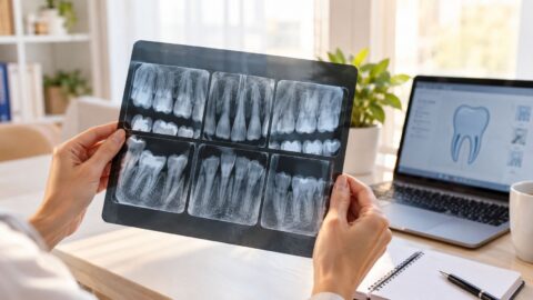
How to Read Dental X Rays with Confidence at Home
You've just finished a cleaning and exam at a Las Vegas dental office, and a screen fills with black, white, and gray shapes. One tooth seems to have a dark shadow. Another looks unusually bright. You may be wondering whether you're seeing a cavity, an old filling, or normal anatomy.
Learning how to read dental X-rays can help you follow your dentist's explanation and ask better questions. It can't replace a clinical exam, a diagnosis, or a treatment plan. If you're searching for a dentist near me, a dentist in Las Vegas, NV, or an emergency dentist for pain or swelling, use an X-ray as part of a conversation with a qualified dental team, not as a stand-alone diagnosis.
Understanding Your Dental X Rays at Aspiring Smiles in Las Vegas
A new patient named Maria recently looked at her digital dental X-rays after an exam in Las Vegas. She noticed several dark spaces around her teeth and immediately worried that each one represented decay. Her dentist slowed down, identified the image type, explained the normal anatomy, and showed her why some dark areas were expected while another shadow needed closer evaluation.
That chairside conversation is the right model for learning. An X-ray is evidence, not a verdict. The image must be matched with symptoms, visual findings, probing, previous films, restorations, and the reason the image was taken. A dark area can represent air, a normal pulp space, or a possible cavity. The same visual feature can mean something different depending on its location and the quality of the image.
Aspiring Smiles Dental and Braces serves families in Las Vegas, NV, including Desert Shores, Sunhampton, Sun City Summerlin, Monterrey, Lone Mountain, Mar-A-Lago, and Painted Desert Estates. The practice describes its care around gentle communication, modern technology, and patient education. Patients who want to understand the technology behind their exam can learn more about advanced dental technology at Aspiring Smiles.
What this guide can and cannot do
You can use this guide to understand:
- The light and dark pattern: Why dense structures appear bright and open spaces appear dark.
- The view type: What bitewing, periapical, and panoramic images are designed to show.
- The reading process: Why dentists check image quality before interpreting a possible finding.
- The next step: When a question calls for routine follow-up or prompt dental care.
You shouldn't use an online image guide to confirm a suspected abscess, decide whether you need a tooth extraction, or plan dental implants near me without an in-person evaluation. For readers who enjoy learning visual diagnostic basics, these beginner-friendly EKG reading tips offer a useful example of why structured observation matters in another field as well.
How Dental X Rays Show Teeth and Bone in Light and Dark
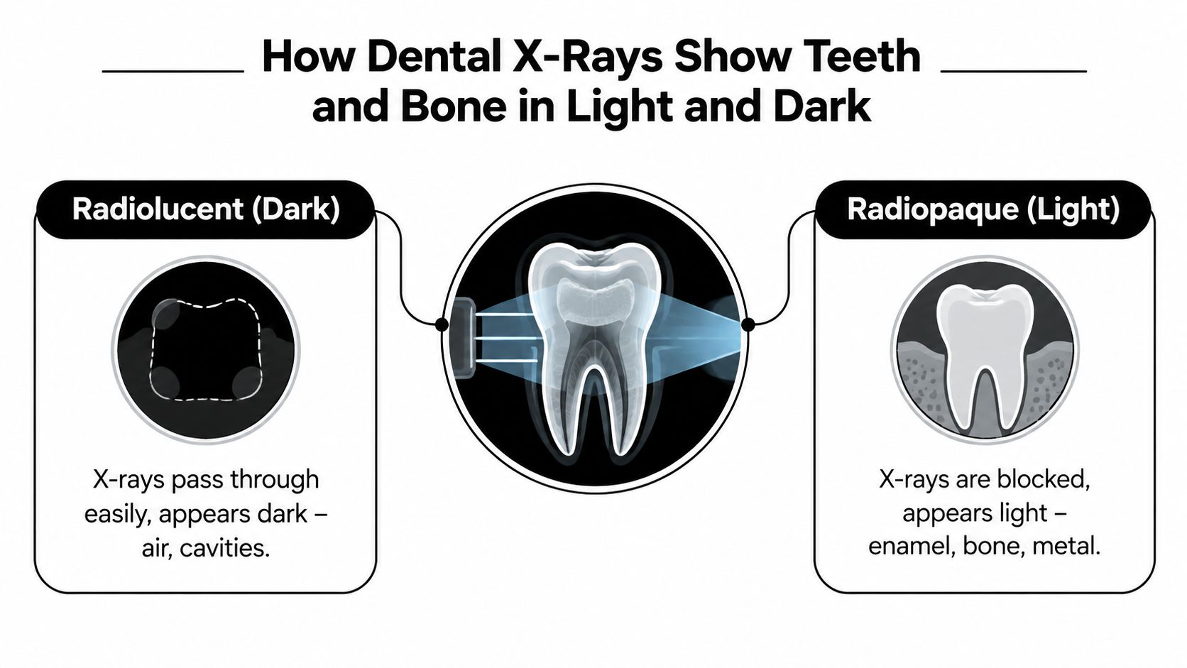
Dental radiographs follow two basic appearance rules. Radiolucent areas look dark because they let more X-rays pass through. Radiopaque areas look white or light gray because they absorb or block more X-rays. This contrast helps a dentist compare one structure with another, but color alone never establishes a diagnosis. (Columbia University dental radiology teaching material)
Start with a normal tooth
On a typical tooth image, enamel forms the brightest natural tooth layer. Dentin is slightly less radiopaque than enamel, while the pulp chamber and root canals appear darker because they contain softer tissue and open spaces. Those boundaries give the dentist a reference for deciding whether a dark change remains in enamel, extends into dentin, or approaches the pulp. (Mustansiriyah University dental anatomy material)
Bone generally appears light gray to white, although its appearance varies with thickness and surrounding anatomy. The thin periodontal ligament space around a tooth root can appear as a narrow dark line. Restorations, crowns, and metal materials usually appear especially bright because they're denser than natural tooth structure.
Use location, not brightness alone
A dark region between two back teeth may deserve attention because that's where the enamel contacts can hide early proximal decay. A dark line inside the root may be the normal canal. A bright area on the biting surface could be a restoration, a dense piece of calculus, or an overlap caused by the angle of the image.
Practical rule: Ask, “Where is this feature, and does it follow normal anatomy?” before asking whether it looks abnormal.
That question keeps you from treating every shadow as a problem. It also explains why your dentist may compare the image with an earlier X-ray or take another view before recommending care.
Bitewing Periapical and Panoramic Views Explained
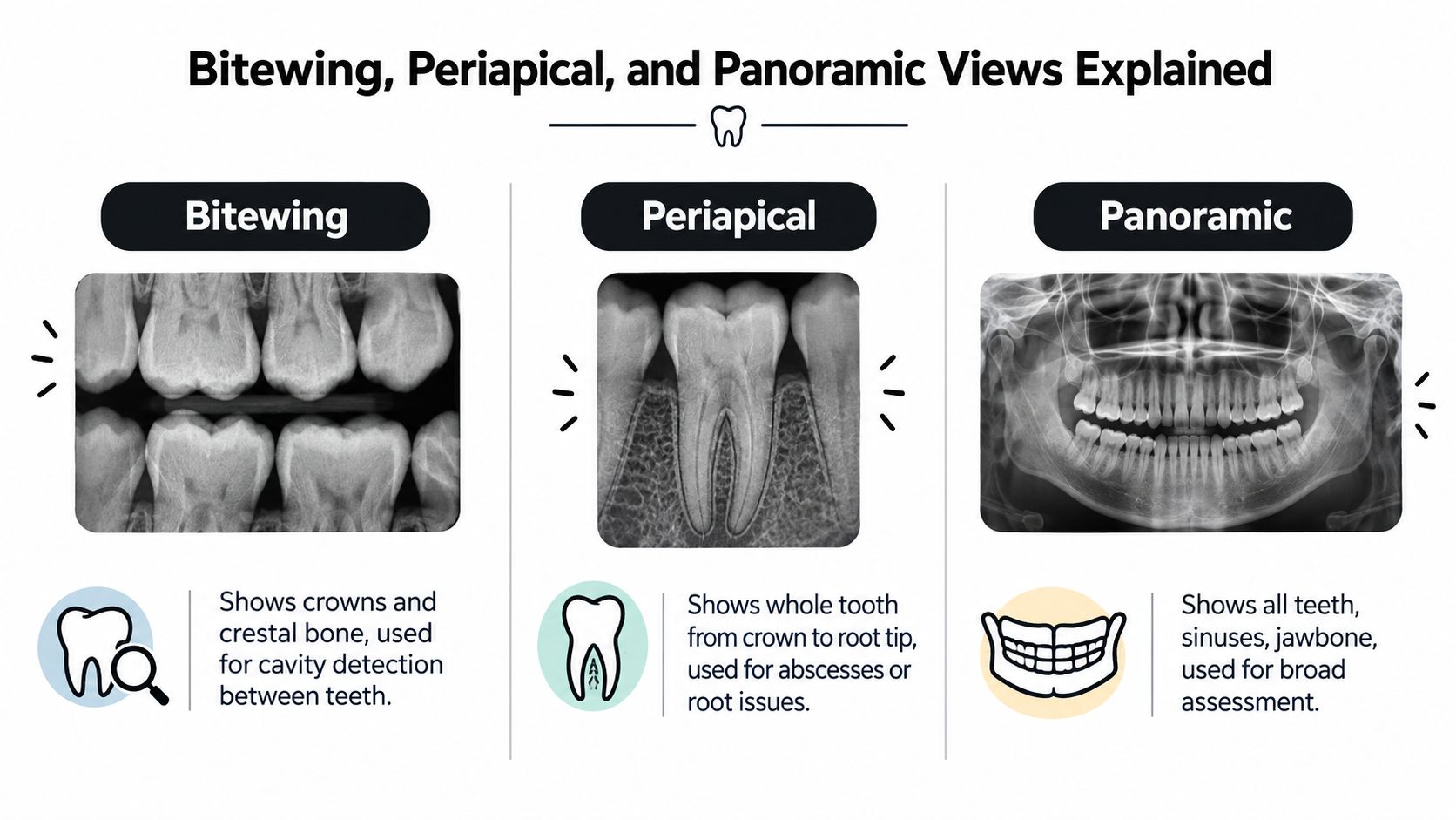
The image type determines the question a dentist can answer. A bitewing concentrates on the crowns of the back teeth and the bone crest between them. A periapical image shows the full length of a selected tooth, including its roots and the surrounding bone near the root tips. A panoramic image provides a broad view of the jaws, teeth, jawbone, and nearby structures such as the sinuses.
Bitewings answer a hidden-cavity question
Bitewings are especially useful when the contact areas between teeth can't be examined directly. They help dentists evaluate interproximal caries, existing restorations, and crestal bone levels. In a study indexed by PubMed, bitewings visualized 79.5% of carious surfaces, compared with 33% for panoramic radiographs, with calculated sensitivity of about 80% versus 33%. (PubMed)
That difference matters because early cavities between teeth can be small and hidden. A panoramic image is valuable for a broad assessment, but it shouldn't be the only image used to screen for early proximal decay.
Periapicals focus on roots and surrounding bone
A dentist may select a periapical image for a painful tooth, suspected root infection, trauma, unusual root anatomy, or a concern near the end of a tooth. Because it includes the root tip, it can help evaluate a dark area around that region when combined with the clinical examination.
Panoramic images provide a wider map
Panoramic radiographs can help assess the overall jaws, wisdom teeth, missing teeth, broad bone patterns, and sinus-related anatomy. They're useful for planning certain treatments, including some evaluations related to dental implants, but their broad view creates less detail for early cavities between neighboring teeth.
The FDA states that posterior bitewing examinations are recommended at 6- to 12-month intervals when proximal surfaces can't be examined visually or with a probe. That's guidance for a particular examination situation, not a universal schedule for every patient. (FDA dental radiographic examination guidance) You can review the practice's dental X-ray services to understand how imaging may fit into a new patient exam.
A Simple Workflow to Check Quality Before You Interpret
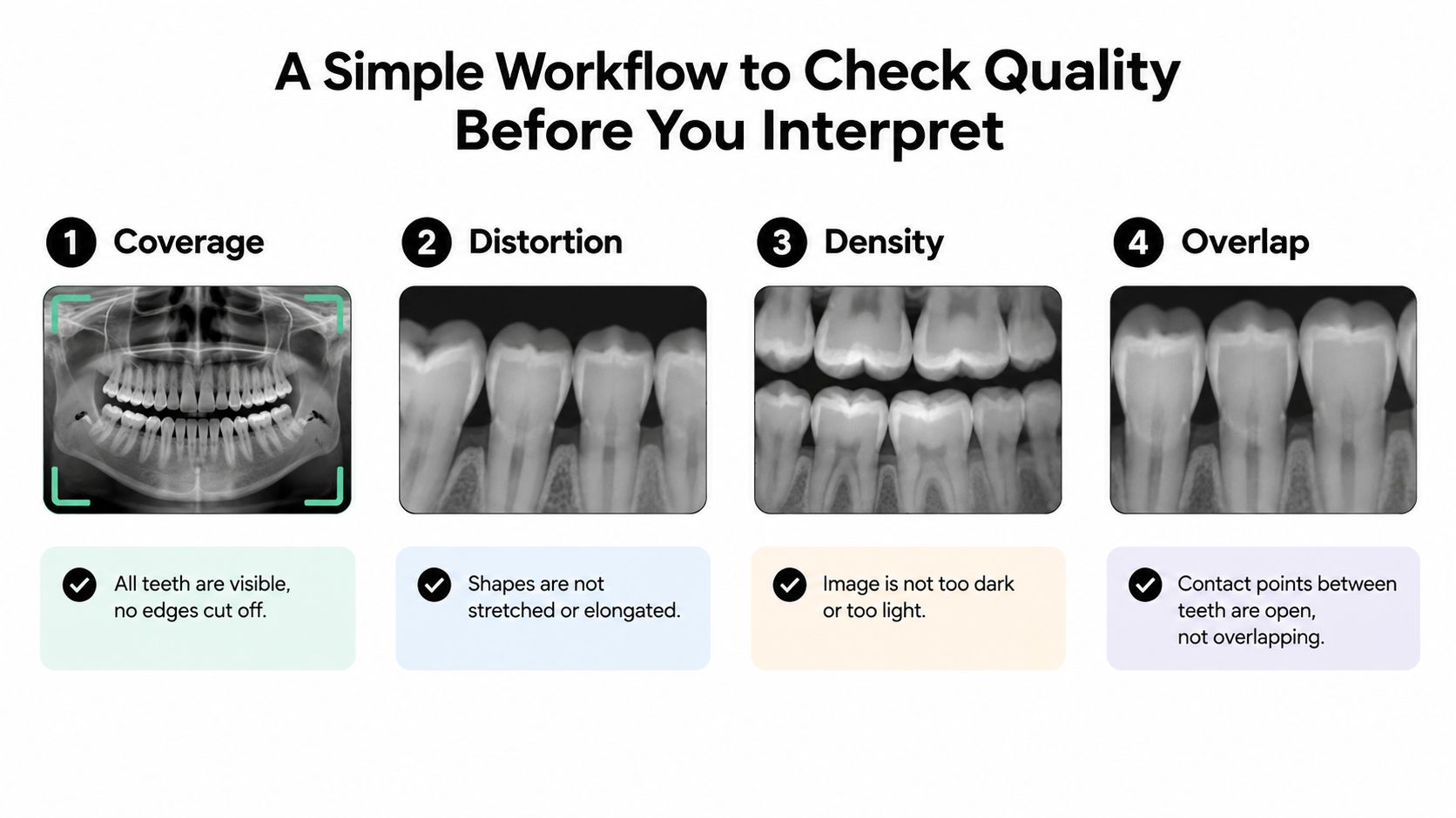
Before you search for a cavity, ask whether the image is good enough to support a conclusion. Coverage, distortion, density, and overlapping contacts can hide a lesion or create a misleading shadow. If the image doesn't capture the needed anatomy, a retake or a different view may be more useful than extended interpretation.
Four quality checks come first
- Coverage: Are the teeth or jaw areas needed for the question visible, or is part of the region cut off?
- Distortion: Do the teeth look stretched, shortened, or unusually angled?
- Density: Is the image light enough and dark enough to show the relevant boundaries?
- Overlap: Are the contact points open enough to inspect, or are neighboring teeth superimposed?
An image can be technically clear and still fail to answer the clinical question. For example, a panoramic film may show the entire mouth but lack the detail needed to rule out a small cavity between two molars. Poor image quality isn't an inconvenience. It's a finding that can limit what a dentist can safely say. (ToothCheck diagnostic image-quality guide)
Move from noticing to diagnosing
Dentists use a stepwise cognitive process:
- Detect: Notice an area that differs from expected anatomy.
- Recognize: Consider what type of finding it may represent.
- Discriminate: Study its edges, location, density, and relationship to nearby structures.
- Diagnose: Combine the image with the exam, symptoms, history, and possible alternatives.
That last step is why reading dental X-rays isn't a single visual glance. A dentist may identify a suspicious area, compare views, test the tooth, and decide whether monitoring, restorative dentistry, root canal treatment, or another service is appropriate.
AI and augmented intelligence are increasingly used to assist with panoramic and periapical interpretation. Review literature indicates that these tools can support performance and decision-making, particularly for less experienced clinicians, while still requiring clinical reasoning and accurate recognition of anatomy versus disease. (PubMed systematic review)
AI can highlight a region for review, but it doesn't replace the dentist who considers pain, bite sensitivity, gum findings, prior treatment, and the quality of the radiograph.
Spotting Normal Anatomy Versus Common Issues on Your Images
Once the image passes a quality check, location and pattern become more useful than isolated color. A small dark notch near a contact between teeth may suggest proximal decay, but overlap, angle, and normal anatomy can create similar appearances. Your dentist will look for a consistent change that crosses expected tooth boundaries and fits the clinical findings.
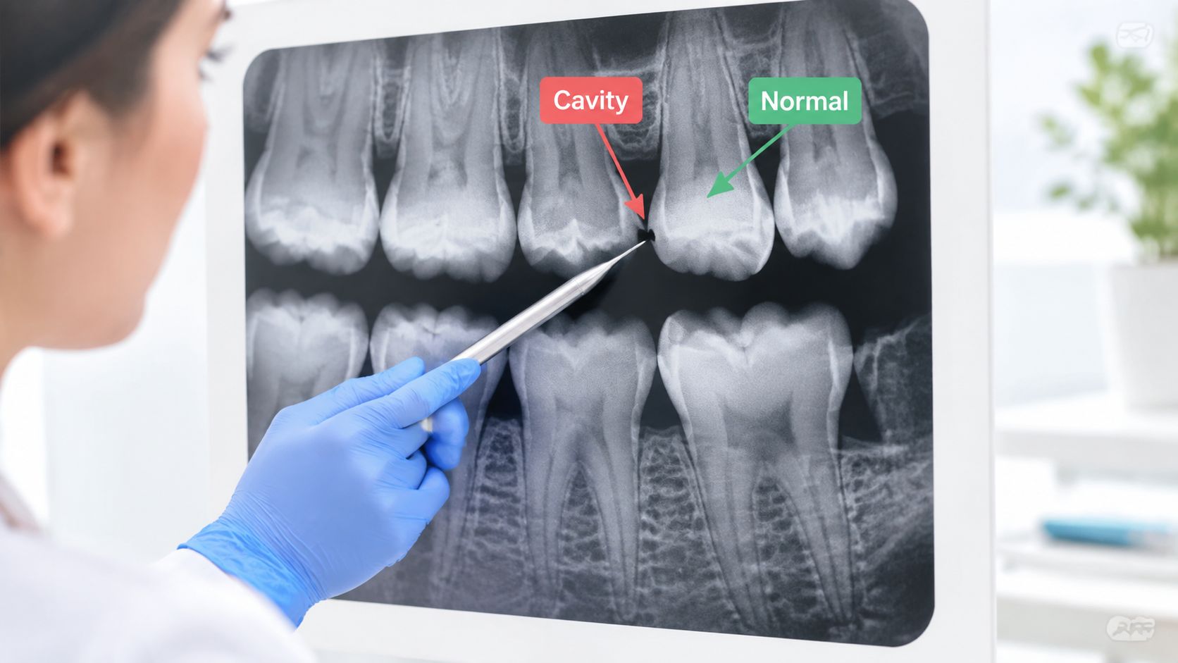
Common patterns worth discussing
A bitewing caries classification describes proximal lesions by how far the radiolucency extends:
- Sound: No radiolucency is visible.
- Initial: The change may reach the dentinoenamel junction or the outer one-third of dentin.
- Moderate: The radiolucency reaches the middle one-third of dentin.
- Extensive: The change reaches the inner one-third of dentin.
A related system labels enamel involvement E1 and E2, and dentin involvement D1 through D3. These labels help clinicians communicate depth, but they don't tell you by themselves whether a tooth needs a filling. The dentist still considers whether the lesion is active, cavitated, accessible for cleaning, and supported by the mouth examination. (Bitewing caries classification review)
Bone, calculus, and root-tip changes
The crest of the bone between teeth normally follows a relatively consistent relationship to the tooth crowns. Lowered bone levels or an uneven, fuzzy crest can raise concern about periodontal changes, especially when paired with bleeding, pocketing, or gum recession. Bright flecks may represent calculus or restorative material, and their meaning depends on shape and location.
A dark halo around a root tip can warrant prompt evaluation because it may be associated with inflammation or infection. It can't confirm an abscess on its own. Pain, facial swelling, fever, drainage, or a tooth injury should prompt contact with an emergency dentist, even if a recent X-ray appears reassuring.
Impacted wisdom teeth and unusual tooth positions are often easier to appreciate on a panoramic view than on bitewings. If a tooth can't be restored, a dentist may discuss tooth extraction and replacement options such as dental implants, but those decisions require an examination and treatment planning. If you want a visual refresher on head and jaw landmarks, you can check out the skull labelling tool from Natomy.
When to Call Your Las Vegas Dentist and What to Expect Next
Call a Las Vegas dental office when an X-ray raises a question, especially if you also have tooth pain, swelling, sensitivity, bleeding gums, a broken restoration, or recent trauma. Don't wait for an online interpretation when symptoms are worsening. A prompt appointment can clarify whether you need preventive care, restorative dentistry, a tooth extraction, or urgent treatment.
Remember: Check image quality first, identify the view type, use light and dark contrasts as clues, and never self-diagnose from an X-ray alone.
At Aspiring Smiles Dental and Braces, located at 3211 N Tenaya Wy Suite 122, patients can expect exams, gentle cleaning and exams, digital dental X-rays, and same-day emergency care when available. Personalized plans may include cosmetic dentistry, teeth whitening, crowns, veneers, Invisalign, restorative care, and dental implants.
The office serves patients across Las Vegas, including Lone Mountain, Painted Desert Estates, and Sun City Summerlin. Flexible payment options, insurance acceptance, and Saturday hours can make it easier to arrange care around work, school, and family schedules. You can request a new patient exam or consultation online when you're ready to discuss your images and next steps.
Aspiring Smiles Dental and Braces provides digital dental X-rays, preventive exams, emergency evaluations, restorative dentistry, cosmetic dentistry, and dental implants for Las Vegas families. If you're looking for a dentist near me and want your X-rays explained clearly, visit Aspiring Smiles Dental and Braces to schedule an appointment or request a consultation.